ITG 500 – Bench AnalyserLaboratory film inspection for samples up to A3
A laboratory bench for film inspection that identifies, classifies and documents optical defects in film samples – in A4 or A3 format.
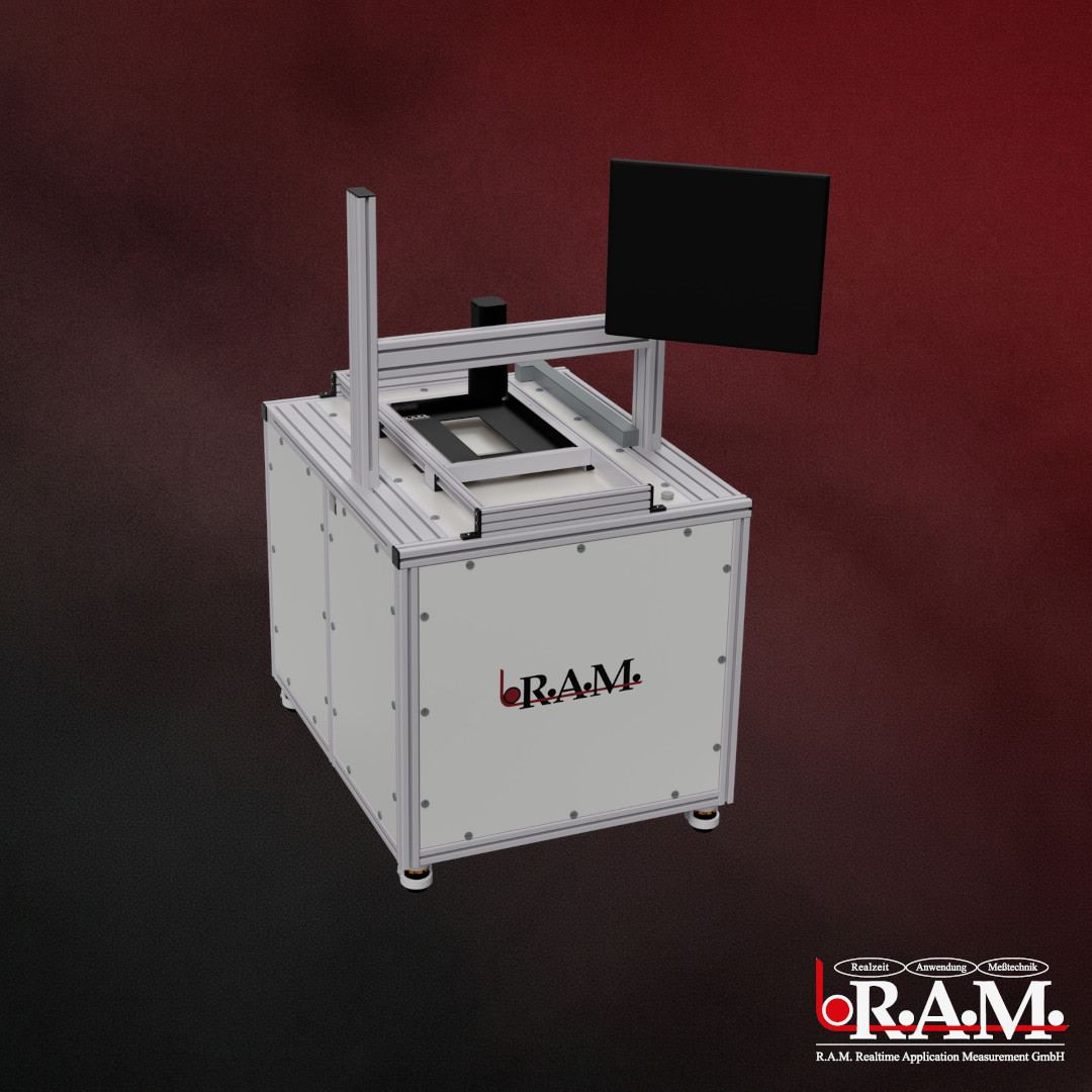 // ITG 500 · Bench Analyser
Precision in lab surface inspection
The Bench Analyser is a laboratory bench for the optical inspection of film samples. It detects, classifies and documents defects in transparent and opaque films – reproducibly and with an image of every defect.
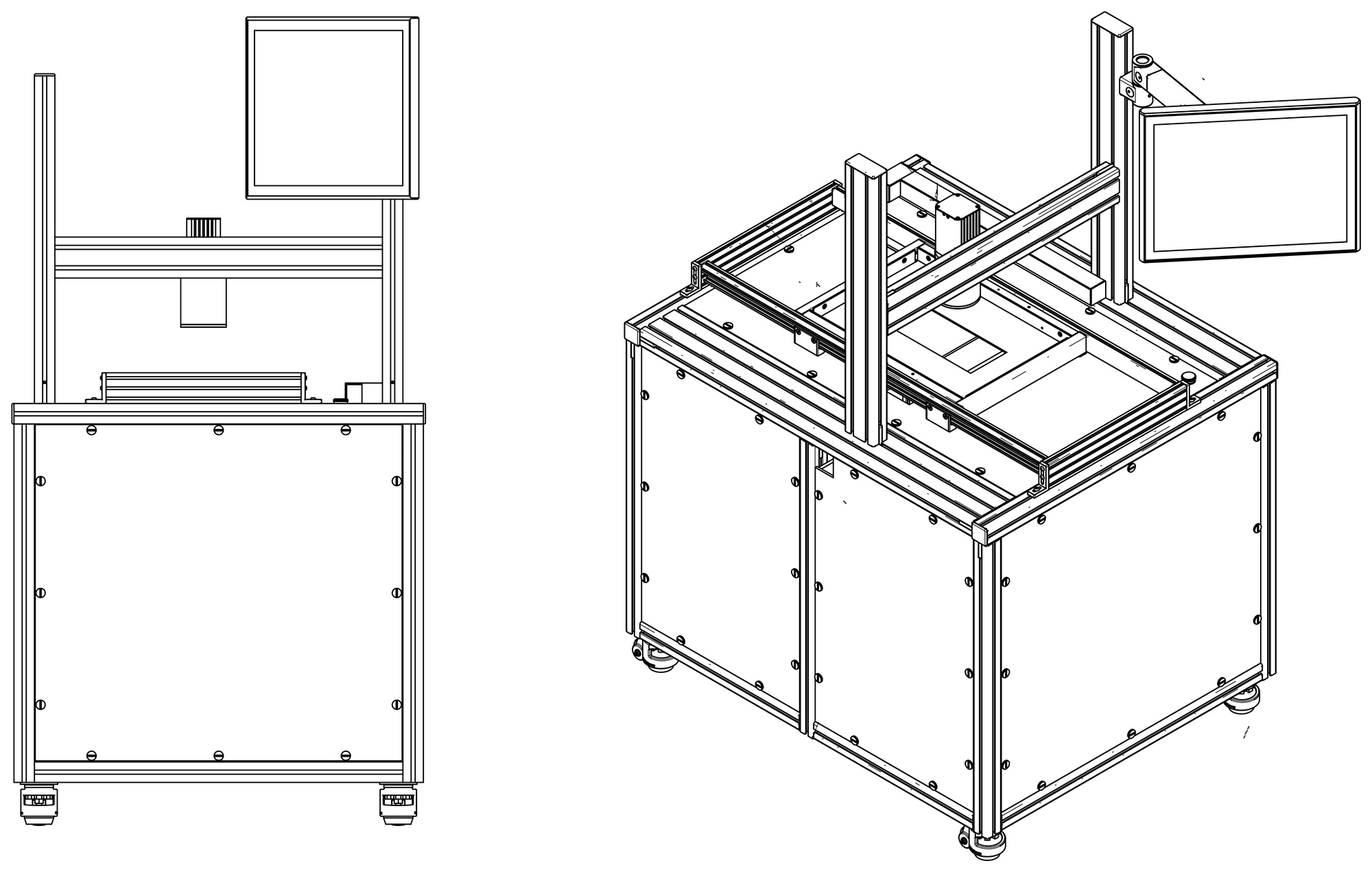
Layout of the laboratory bench
- 1CameraLine camera based on GigE Vision (4K) – captures the sample line by line from above.
- 2Carriage and sample bedHold the film sample up to DIN A3 and move it beneath the camera.
- 3Touch monitor on swivel armOperation via CAMEN with on-screen keyboard; mouse and keyboard can be connected.
- 4Opening for dark-field apertureHolds the dark-field aperture – for gel specks in transparent films.
- 5Castors for mobilityThe laboratory bench can be moved flexibly within the lab.
Four illumination modes
Transmitted light
For transparent films – with white and opaque films the contrast is often insufficient.
Reflected light
For white, opaque, metallised and coated films – reveals surface defects.
Dark-field aperture
Specifically designed to detect gel specks in transparent films.
Dual illumination
Transmitted and reflected light combined.
Automatically detected and classified
From gel specks, gels and burners through fisheyes to impurities, wrinkles, voids or flies – the system classifies them all and additionally provides photos of each defect for detailed evaluation.
Gel specks
Tiny gel points in transparent films.
Gels
Gel inclusions in the material.
Burn marks
Burnt particles and dark spots.
Fisheyes
Lens-shaped defects.
Impurities
Foreign particles and inclusions.
Wrinkles
Wrinkles and creases in the film.
Voids
Holes and voids.
Flies
Small fly-like specks.
Five steps to a result
The bench analyser is operated through CAMEN – on the touchscreen, with entries made via an on-screen keyboard. A mouse and keyboard can be connected.
Place the sample
Place the film sample flat and free of wrinkles on the carriage’s support surface. For transparent films in dark field, insert the dark-field aperture.
Load recipe
All product-related settings sit in the recipe. Loading it sets the camera automatically – so keep one recipe per material.
Calibrate
Calibrate the camera with the sample in place: exposure time, gain, grey value. With the opacity option, opacity is zeroed beforehand without material.
Scan
The sample is scanned. Defects are counted immediately and placed on the defect map at their actual position.
Roll report
CAMEN creates the roll report with all defects and defect images; job data such as job number, customer and sample name are included.
You decide what gets counted
Size and type classifiers can be combined freely. Each class is given a name, a symbol and a colour – exactly how the defect will later appear on the defect map.
Size classifiers
Classes defined by minimum and maximum size. Quick to set up, but limited in how finely they discriminate.
Type classifiers
Taken from a trained classifier list. They take numerous defect properties into account and therefore discriminate far more finely.
Templates
Classifier lists can be saved as templates and – unlike recipes – exchanged between systems. Two standard templates are supplied: three types with three or four size classes.
Basic types
Dark, darkest, light, lightest and combined. Combined defects are enclosed defects – for instance a dark rim with a hole in the middle.
Warning and alarm limits
Per classifier, for the whole roll or per segment. Warning values appear in yellow, alarm values in red.
Defect counter
Counts per classifier – either in absolute terms or related to area (1/100 cm²).
Three display modes
The defect map shows size symbols, defect images, or both together.
Connection to your network
Network integration
Fits into your IT network – accessible from any workstation.
Real-time evaluation
Monitor evaluations in real time.
USB data export
Download data directly via the USB interface.
Specifications
Subject to technical changes. Details of your configuration are available on request.
RelatedSample analysis in our laboratory in Flörsheim · Service, maintenance and remote support · Training for operators, production leads and maintenance
Frequently asked questions about the ITG 500 – Bench Analyser
Your question isn’t here? Just reach out.
Film samples in A4 or A3 format – larger than the 180 mm or 100 mm measuring window –, transparent, white, opaque, metallised and coated films. The right illumination is selected for each material and saved in the recipe.
Transmitted light suits transparent films, the dark-field aperture very low-contrast defects such as fine gel specks. For white, opaque, metallised and coated films, reflected light provides better contrast.
With the 4K line-scan camera approx. 60 µm; the measuring window is 180 mm or 100 mm. Cameras with 2K, 6K, 8K or 12K are available for other resolutions.
Inspection runs in CAMEN; reports, defect images and statistics are available via vSTAT – stored locally in the internal SQL database. Would you like to test your material first? We carry out the sample analysis in our laboratory.
Interested in the ITG 500 – Bench Analyser?
We will show you how the Bench Analyser can advance your quality lab and production.